- About BNMS
- HIstory of BNMS
- BNMS People
- BNMS Policies
- BNMS Achievements
- Governance
- Awards & Prizes
- Donate/Support Us
- About Nuclear Medicine
- FAQs
- Membership
- Events
- News
- Resources
- Radionuclide Supply
- UKRG
- MRT Consortium
| FDG PET Scan |
|
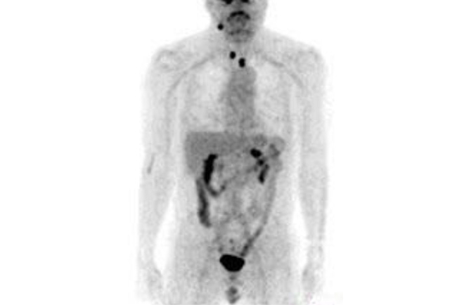
What is an FDG PET scan? PET is short for Positron Emission Tomography. It is a nuclear medicine technique that uses a radioactive substance to look at different parts of the body in a unique way. PET scans can be performed for a variety of different conditions. An FDG PET scan is the most common type. It involves an injection into a vein in your arm of FDG, a substance similar to glucose. The PET scanner then detects areas of high glucose concentration within the body.
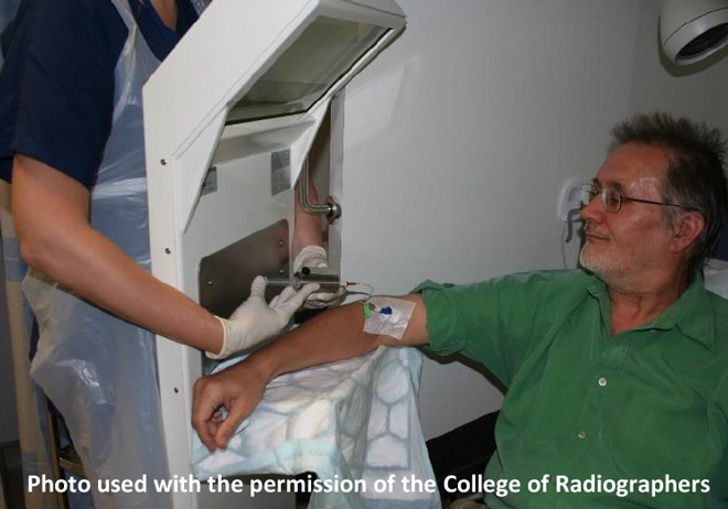
Is it safe for me to have the scan? For this scan it is necessary to inject a small amount of radioactive tracer called FDG in order to take the pictures. The small risk from this (similar to a CT scan) is outweighed by the information that will be gained by taking the scan. A doctor will have checked the request to make sure this is the appropriate test for you. If you have any concerns or would like further information, please contact the department where you are having your PET scan. If you don’t understand why you need to have this scan, please speak to the doctor who referred you. For female patients aged 12 to 55 If you know that you are pregnant, or there is any chance that you might be pregnant, please contact the department where you will be having your scan. Do this as soon as possible as the scan can be postponed if it is not urgent. Also, contact the department if you are breast-feeding, as they may give you special instructions. Preparation for your scan It is important that you follow instructions to do with fasting before your scan. Please drink plain unflavoured water but nothing else during the fasting period. If you are taking any liquid medicine please contact the nuclear medicine department for advice, as it may contain sugar. Unless your appointment letter says otherwise, you can take any tablets or capsules as normal. You should also avoid any strenuous exercise for 12 hours before your appointment. If you are diabetic, it is also really important to let the department know as soon as possible before your appointment. They will discuss how you can control your diabetes. They will give you specific advice about when to eat and when to take any medication in order to make sure that your blood sugar is in the correct range when The FDG will be ordered as a timed delivery especially for you. Please do not be late for your appointment or you may not be able to have your scan that day. When you arrive In order to make sure that your scan is of the best quality, your blood sugar level will be tested when you arrive. If your sugar level is too high it may not be possible to do the scan. Your injection The FDG is injected through a short tube connected to a needle in your arm. You may have had a blood test in the past; this is much the same. The ‘pinprick’ of the needle may hurt a bit, but that is all.
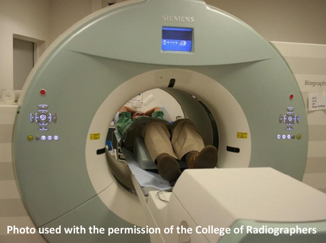
Your scan It takes a while for sufficient FDG to be taken up by the cells in your body, so after the injection you will usually be asked to wait for about 1 hour before the scan is taken. During this waiting time you will be asked to rest in a quiet area. This will help to improve the quality of your scan. The pictures will be taken by a machine called a PET scanner. It has a short tunnel that you will have to The scanner will also take some additional images using an X-ray CT scanner, but these only take a few One of the clinical staff will be watching you from behind a screen all the time to make sure that you are OK.
After your scan It is very unlikely that you will feel any side-effects after the scan, but if you think that you do, please let the nuclear medicine department know. You may continue all your normal activities unless you have been advised otherwise. After your scan there will be some radioactivity left in your body but this will not present a significant risk to other people around you. However, for the rest of the day, we suggest that you try to keep any time that you spend within arm’s length of pregnant women, babies and small children as short as possible; but there is no need The radioactivity in your body will soon disappear, but if you drink plenty of fluids this will help to clear the radioactivity more quickly. Travelling abroad It is perfectly safe for you to travel abroad after your scan, but many airports and sea ports are now equipped with very sensitive radiation detectors. So it is possible that the very small amount of radioactivity left in your body could set off a detector as you pass through security. Therefore, if you intend to travel abroad within 3 days Your results Your PET scan will be looked at by a specialist who will issue a report. The report will be sent to the doctor who requested your scan rather than to your GP. This is because the doctor who requested your scan will have all the results from other tests and will be able to tell you how the result of your PET scan affects your care. Information about you As part of your care, information will be shared between clinical staff, some of whom you may not meet. It may also be used to help train other staff. Information collected may also be used later on to help the department improve their quality of care, plan services or to research into new developments. The pictures from your scan may be used to teach other healthcare workers, but your name and all other identification will be removed first. It won’t be possible to identify you from the scan pictures. All information will be treated as confidential and is not given to anyone who does not need it. If you have any concerns, please discuss these with the department. More information All the staff would like to make your visit as pleasant as possible. If you have any concerns please talk to a member of the nuclear medicine staff. This video, made in the Central Manchester Nuclear Medicine Centre, shows a patient having a PET Scan.
A printable version of this leaflet can be found here |
19 hours agoWavelength - July 2026
30/06/2026Wavelength - June 2026
15/09/2026
BNMS MRT Webinar Series - Current radionuclide therapy service delivery from a physicist’s perspecti
18/09/2026 » 20/09/2026
ACNM Annual Meeting
 BNMS YouTube Channel #BNMS2026 https://www.bnms.org.uk/news/721432/NEW-Feature-Member.htm
 RCR NM Travelling Professor https://www.bnms.org.uk/news/728160/BNMS-Feature-Member.htm